The human eye is a complex organ enabling vision by capturing and processing light. It functions like a sophisticated camera, with precise parts working together for sharp images.
Key Parts
The eye has three main layers: the outer fibrous tunic (cornea and sclera), middle vascular tunic (choroid, ciliary body, iris), and inner retina. External structures include the sclera (white protective layer), conjunctiva (moist lining), cornea (clear front dome), iris (colored muscle), and pupil (light aperture). Internal parts feature the lens (focuses light), aqueous humor (front fluid), vitreous humor (rear gel), macula (central vision spot), fovea (sharp focus area), optic disc (nerve exit), and optic nerve (signal transmitter).
How Vision Works
Light enters through the cornea, which refracts most of it, then passes the pupil (iris-controlled) and lens for fine focusing onto the retina. Retina cells—rods for low light/motion and cones for color/detail—convert light to electrical signals sent via the optic nerve to the brain. Fluids like aqueous and vitreous humor maintain shape and nourishment.
Unique Characteristics
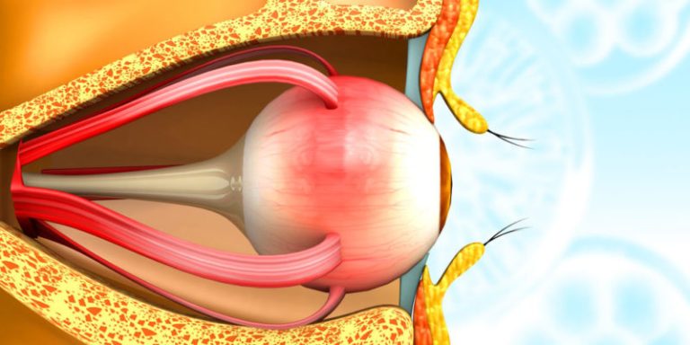
Humans have two forward-facing eyes in bony orbits, moved by six extraocular muscles for binocular depth perception. The cornea lacks blood vessels for clarity, deriving oxygen from air/tears. Cones peak at the fovea for 6-7 million high-resolution cells, while rods enable night vision. The eye adjusts focus via ciliary muscles (accommodation) and adapts to light via pupil/iris. It processes ~10 million bits of visual data per second, with the brain inverting upside-down retinal images.