If you have ever wondered how a beam of light bouncing off a distant mountain, a printed page, or a loved one’s face becomes a picture in your mind, you have already started asking the central question of ophthalmology. In more than a decade of examining eyes across every age group — from newborns undergoing their first red-reflex test to patients navigating cataract surgery in their eighties — I have come to see the human eye not just as an organ, but as the most elegant piece of biological engineering most of us never stop to appreciate.
This guide walks through the human eye the way I would explain it to a patient sitting across from me in the exam chair: what it is, how it is built, how each part earns its place, how the whole system works together to produce sight, and what makes this organ so remarkable — and so vulnerable. Whether you are a student, a caregiver, or simply someone who wants to understand the two organs you rely on every waking moment, this article is written to give you a genuinely thorough, medically grounded understanding of the human eye.
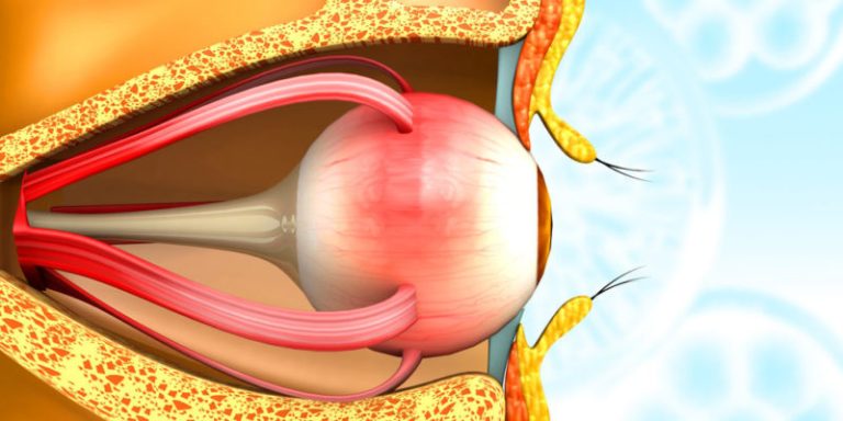
What Is the Human Eye?
The human eye is a specialized sensory organ that converts light into electrical signals the brain can interpret as vision. Roughly spherical and about 24 millimeters in diameter — close to the size of a large marble — each eye weighs approximately 7.5 grams, yet it contains more than two million working parts, including muscles, nerves, and light-sensitive cells.
Anatomically, the eye is often compared to a camera, and while the analogy is imperfect, it is a useful starting point. Light enters through a clear front window (the cornea), passes through an adjustable aperture (the pupil), is focused by an internal lens, and lands on a light-sensitive “film” at the back (the retina). But unlike a camera, the eye does not simply capture a static image — it continuously adjusts focus, light intake, and even color sensitivity in real time, and it hands its raw data to a brain that reconstructs a three-dimensional, color-corrected, motion-stabilized picture of the world dozens of times per second.
The eye does not work in isolation. It is part of the broader visual system, which includes the optic nerve, the optic pathways inside the brain, and the visual cortex at the back of the skull. Understanding the eye, then, is really the first half of understanding how human beings see — the second half happens entirely inside the brain.
Why Understanding Eye Anatomy Matters
Patients frequently ask me why they should care about the technical names of eye structures. The honest answer: because nearly every eye complaint — blurry vision, redness, floaters, glare, double vision — traces back to a specific part of this anatomy doing something unexpected. Knowing the parts of the eye and what each one does gives you a framework for understanding your own eye health, asking better questions during checkups, and recognizing warning signs early, when they are most treatable.
The Layered Structure of the Eyeball
Before looking at individual parts, it helps to understand that the eyeball itself is built from three concentric layers, or “coats,” much like the layers of an onion.
The Fibrous Layer (Sclera and Cornea)
The outermost layer is tough and protective. Most of it is the sclera — the white part of the eye — made of dense, interwoven collagen fibers that give the eyeball its shape and shield it from injury. At the very front, this fibrous layer becomes transparent and curves outward slightly to form the cornea, the eye’s primary focusing surface.
The Vascular Layer (Uvea)
The middle layer, called the uvea, is rich in blood vessels and pigment. It consists of three connected structures: the choroid at the back, the ciliary body in the middle, and the iris at the front. This layer nourishes the eye and controls light entry and lens focusing, as described in more detail below.
The Neural Layer (Retina)
The innermost layer is the retina, a thin sheet of light-sensitive nervous tissue that lines the back two-thirds of the eyeball. This is where light is actually converted into the electrical signals that become vision.
With this three-layer framework in mind, let’s walk through the eye’s individual parts, front to back — the same “tour” I use when I explain the anatomy to my own patients.
The External and Protective Structures of the Eye
Eyelids and Eyelashes
The eyelids are the eye’s first line of defense. Every blink — and we blink roughly 15 to 20 times per minute — spreads a thin film of tears across the eye’s surface, clearing away dust and debris and keeping the cornea moist. The eyelashes lining the lid margins act as a sensory tripwire, triggering an automatic blink reflex when something approaches the eye too closely.
Conjunctiva
The conjunctiva is a thin, transparent mucous membrane that lines the inside of the eyelids and covers the white of the eye (but not the cornea). It contains tiny blood vessels and mucus-producing cells that help lubricate the eye. When this membrane becomes inflamed — a condition most people know as “pink eye” — the eye turns red and irritated, which is one of the most common reasons patients come to see me.
Tear Film and Lacrimal Glands
Sitting above and to the outer side of each eye, the lacrimal glands continuously produce tears, which are far more sophisticated than plain water. The tear film has three layers — an oily outer layer that slows evaporation, a watery middle layer that provides nutrients and washes away debris, and a mucous inner layer that helps the tears stick evenly to the eye’s surface. An imbalance in any of these layers is what causes dry eye syndrome, one of the most frequent complaints I see in clinic, especially among people who spend long hours looking at screens.
Sclera
Already introduced as part of the fibrous outer layer, the sclera is the tough, white, opaque tissue that makes up about five-sixths of the eyeball’s outer surface. Six extraocular muscles attach directly to the sclera, allowing the eye to move up, down, side to side, and rotate. Beyond its protective role, the sclera’s rigidity also helps maintain the eye’s precise internal pressure and shape, both of which are essential for clear focusing.
The Front-of-Eye Structures: Where Light First Enters
Cornea
The cornea is the clear, dome-shaped front surface of the eye, and it is arguably the single most powerful focusing structure you have. Although it looks like a simple transparent window, the cornea actually contributes roughly two-thirds of the eye’s total focusing power — more than the internal lens. It is also one of the only tissues in the body with no blood vessels; it receives oxygen and nutrients directly from tears and the fluid behind it, which is part of why it heals slowly compared to other tissues and why corneal injuries and infections need prompt attention.
The cornea has five distinct microscopic layers — epithelium, Bowman’s layer, stroma, Descemet’s membrane, and endothelium — each with a specialized job in maintaining clarity and structural strength. Irregularities in corneal curvature are what cause astigmatism, one of the most common refractive errors I diagnose.
Anterior Chamber and Aqueous Humor
Just behind the cornea lies the anterior chamber, a fluid-filled space containing a clear liquid called aqueous humor. This fluid is constantly produced by the ciliary body, circulates through the pupil, and drains out through a mesh-like structure called the trabecular meshwork. This continuous production-and-drainage cycle maintains intraocular pressure — the pressure inside the eye. When drainage becomes blocked or inefficient, pressure builds up, which is the underlying mechanism behind glaucoma, a leading cause of irreversible blindness worldwide.
Iris
The iris is the colored, ring-shaped structure you see when you look at someone’s eyes — brown, blue, green, hazel, or gray. Its color comes from the amount and type of pigment (melanin) in its tissue; more melanin produces darker irises, while less melanin allows blue light-scattering effects to dominate, producing lighter colors. Functionally, the iris acts like the aperture ring on a camera lens. It contains two sets of tiny muscles: the sphincter muscle, which constricts the pupil in bright light, and the dilator muscle, which widens it in dim light. This automatic light-regulating reflex protects the retina from overexposure while ensuring enough light reaches it in darker settings.
Pupil
The pupil is not a physical structure but rather the open, circular space in the center of the iris through which light passes. It appears black because most light entering the eye is absorbed by the tissues inside rather than reflected back out. Pupil size typically ranges from about 2 millimeters in bright light to 8 millimeters in near-total darkness, and this reflex, controlled by the autonomic nervous system, is so reliable that abnormal pupil responses are one of the first things I check when evaluating for neurological problems.
Lens
Positioned directly behind the iris and held in place by a ring of fine fibers called zonules, the crystalline lens is a flexible, transparent, biconvex structure that fine-tunes focus after the cornea has done the bulk of the work. Unlike the cornea, the lens can change shape through a process called accommodation: the ciliary muscles contract to make the lens rounder for near vision and relax to flatten it for distance vision. This is the same mechanism that gradually stiffens with age, leading to presbyopia — the reason most people need reading glasses by their mid-forties, even if they never needed glasses before. The lens is also where cataracts form, as proteins within it become cloudy over decades of UV exposure and normal aging.
Ciliary Body
Often overlooked by patients but essential to eye function, the ciliary body sits just behind the iris and performs two jobs at once: it produces aqueous humor, and its ciliary muscles control the shape of the lens during accommodation. It is a small structure with an outsized influence on both eye pressure and focusing ability.
The Internal Chamber and Middle Structures
Vitreous Humor and the Vitreous Cavity
Behind the lens lies the largest space in the eye, the vitreous cavity, filled with a clear, gel-like substance called the vitreous humor. Made mostly of water, collagen, and hyaluronic acid, the vitreous gives the eyeball its round shape and helps keep the retina pressed firmly against the back wall of the eye. With age, the vitreous gradually liquefies and can pull away from the retina, a normal process that often produces the floating spots or “floaters” many people notice, especially against a bright sky or blank wall. While usually harmless, a sudden shower of new floaters accompanied by flashes of light warrants urgent evaluation, since it can signal a retinal tear.
Choroid
Sandwiched between the sclera and the retina, the choroid is a thin, richly vascular layer that supplies oxygen and nutrients to the outer retina, particularly the photoreceptor cells. Its dense network of blood vessels also gives it a dark pigmentation that helps absorb stray light within the eye, reducing internal glare and improving image clarity — much like the matte black interior of a camera body.
The Retina and Optic Nerve: Where Sight Truly Begins
Retina
The retina is, in many ways, the most important real estate in the entire visual system. This thin, multilayered sheet of neural tissue lines the back of the eye and contains over 120 million photoreceptor cells of two main types:
- Rods, numbering around 120 million per eye, are extremely sensitive to low light and are responsible for night vision and peripheral vision, though they cannot distinguish color.
- Cones, numbering around 6 million per eye, function best in bright light and are responsible for color vision and fine visual detail. Humans have three types of cones, each sensitive to different wavelengths of light (roughly corresponding to red, green, and blue), and it is the combined signal from these three cone types that produces the full spectrum of color we perceive.
When light strikes these photoreceptors, it triggers a biochemical cascade called phototransduction, converting light energy into an electrical nerve signal. These signals pass through several layers of retinal neurons — bipolar cells, then ganglion cells — before leaving the eye altogether.
Macula and Fovea
Near the center of the retina lies the macula, a small, specialized region responsible for sharp, detailed central vision — the vision you use for reading, recognizing faces, and driving. At the very center of the macula is the fovea, a tiny pit densely packed with cones and virtually no rods, making it the point of highest visual acuity in the entire eye. Degeneration of this region, known as macular degeneration, is one of the leading causes of vision loss in people over 60, and it is a condition I screen for carefully during routine dilated exams.
Optic Disc and Optic Nerve
All the signals gathered by the retina’s roughly 1.2 million ganglion cells converge and exit the eye through a single point called the optic disc, forming the optic nerve. Because this point contains no photoreceptors, it corresponds to a natural blind spot in each eye’s visual field — one that you never notice because your brain fills in the gap using information from the other eye and surrounding context. The optic nerve then carries these electrical signals along a complex pathway through the optic chiasm, where fibers from each eye partially cross over, and on to the visual cortex at the back of the brain, where the raw signals are finally assembled into the conscious experience of seeing.
How the Human Eye Actually Produces Vision: Step by Step
Understanding the individual parts is only half the picture. What makes the eye remarkable is how seamlessly these parts cooperate, many times per second, without any conscious effort on your part. Here is the sequence, in the order it actually happens:
- Light enters the eye through the cornea, which performs the majority of the initial focusing.
- The iris adjusts pupil size almost instantly, controlling how much light is allowed through based on ambient brightness.
- The lens fine-tunes focus through accommodation, changing shape depending on whether you are looking at something near or far.
- Light travels through the vitreous humor and lands on the retina at the back of the eye.
- Photoreceptor cells convert light into electrical signals through phototransduction, with rods handling dim light and motion, and cones handling color and detail.
- Signals are processed within retinal layers, compressing and organizing the information before it leaves the eye.
- The optic nerve transmits signals to the brain, where the visual cortex interprets them — correcting for the fact that the image projected on the retina is actually upside down and reversed, combining input from both eyes into a single three-dimensional image, and stabilizing the picture despite constant small eye movements.
This entire chain, from light hitting the cornea to conscious perception in the brain, takes a matter of milliseconds — and it happens continuously, tens of times per second, for every waking moment of your life.
Binocular Vision and Depth Perception
One of the most functionally important characteristics of the human visual system is that we have two eyes positioned a few centimeters apart, each capturing a slightly different view of the world. The brain compares these two slightly offset images — a process called stereopsis — to calculate depth and distance with remarkable precision. This is why closing one eye makes it noticeably harder to judge how far away an object is, thread a needle, or catch a ball. Binocular vision also expands the total field of view to roughly 200 degrees horizontally, with a central overlapping zone of about 120 degrees where true depth perception occurs.
Color Vision and Light Sensitivity
Human color vision, made possible by the three types of cone cells described earlier, allows most people to distinguish roughly one million different shades and hues. This trichromatic system is a defining characteristic of human sight, though it is not universal — some people are born with a deficiency in one or more cone types, resulting in color vision deficiency, commonly called color blindness, which affects an estimated 1 in 12 men and 1 in 200 women, primarily involving difficulty distinguishing red and green.
The eye’s sensitivity to light intensity is equally impressive. Through a combination of pupil constriction and a biochemical process called dark adaptation — in which the retina’s photopigments regenerate and become more sensitive over 20 to 30 minutes in darkness — human eyes can function across a light range spanning roughly ten orders of magnitude, from bright sunlight to near-total darkness.
Key Characteristics That Make the Human Eye Unique
A few defining traits set the human eye apart, and they are worth understanding on their own:
- Self-focusing optics: The combination of a fixed-curvature cornea and an adjustable lens gives the eye both power and flexibility — a hybrid optical system no consumer camera fully replicates.
- Continuous self-lubrication: The tear film is refreshed with every blink, and the eye maintains its own moisture, clarity, and mild antimicrobial protection through enzymes like lysozyme found in tears.
- Self-regulating light control: The iris and retina work together to allow functional vision across an enormous range of lighting conditions, adjusting automatically without conscious input.
- A privileged immune status: The eye, particularly the cornea and the front chamber, has what is called “immune privilege” — a dampened immune response that reduces inflammation and protects delicate, irreplaceable tissue from collateral damage during infection, at some cost to how aggressively it can fight invaders.
- Minimal regenerative capacity: Unlike skin or liver tissue, retinal neurons and optic nerve fibers generally do not regenerate once damaged, which is precisely why conditions like glaucoma and optic nerve injury cause permanent vision loss and why early detection is so clinically important.
- Constant micro-movement: Even when you believe you are staring at something perfectly still, your eyes make tiny involuntary movements called microsaccades, which prevent photoreceptors from adapting to a static image and going “blind” to unchanging detail.
Common Conditions That Affect the Human Eye
Because nearly every structure described above can be affected by disease, aging, or injury, it is worth briefly connecting anatomy to the conditions I see most often in practice:
- Refractive errors (myopia, hyperopia, astigmatism, presbyopia) arise from mismatches between the eye’s focusing power and its length or corneal curvature, or from age-related lens stiffening.
- Cataracts develop when proteins in the lens clump together and cloud over time, most commonly due to aging and cumulative UV exposure.
- Glaucoma results from elevated intraocular pressure damaging the optic nerve, often silently, which is why routine eye pressure checks matter even without symptoms.
- Macular degeneration involves deterioration of the macula, affecting central vision while typically sparing peripheral sight.
- Dry eye syndrome stems from an imbalance in tear film production or composition.
- Diabetic retinopathy damages the delicate blood vessels of the retina, making it a leading cause of vision loss among working-age adults with diabetes.
- Conjunctivitis (pink eye) involves inflammation of the conjunctiva, usually from infection or allergy.
Practical Tips for Protecting Your Eye Health
As someone who spends most working days looking directly into other people’s eyes, I can say confidently that the majority of long-term vision problems are either preventable or far more manageable when caught early. A few habits go a long way:
- Get a comprehensive dilated eye exam regularly — even without symptoms, since many serious conditions like glaucoma and early macular degeneration are silent in their initial stages.
- Wear UV-protective sunglasses outdoors to reduce cumulative damage linked to cataracts and certain retinal conditions.
- Follow the 20-20-20 rule during screen use — every 20 minutes, look at something 20 feet away for at least 20 seconds — to ease digital eye strain and reduce dry eye symptoms.
- Manage systemic health conditions, particularly diabetes and high blood pressure, since both directly affect the delicate blood vessels of the retina.
- Never skip protective eyewear during sports, yard work, or activities involving flying debris or chemicals, since the eye’s own healing capacity is limited.
- Stay hydrated and blink consciously during long periods of screen focus, since concentrated visual tasks reduce blink rate and dry out the tear film.
Frequently Asked Questions About the Human Eye
How many parts does the human eye have?
The eye is typically described in terms of a dozen or so major structures — including the cornea, sclera, iris, pupil, lens, ciliary body, choroid, retina, macula, vitreous humor, and optic nerve — though at a microscopic level it contains millions of individual cells, each with a specialized role.
Why do eyes come in different colors?
Eye color is determined mainly by the amount and distribution of melanin pigment in the iris, along with how light scatters within its tissue; genetics involving multiple genes determines the final combination.
Can the human eye actually see in the dark?
Not in complete darkness, but human eyes are remarkably good at adapting to low light through the process of dark adaptation, during which rod photoreceptors become progressively more sensitive over 20 to 30 minutes.
Why do we have a blind spot, and why don’t we notice it?
The blind spot exists where the optic nerve exits the eye, an area with no photoreceptors. We don’t notice it because each eye’s blind spot falls in a different part of the visual field, and the brain fills the gap using surrounding visual information and input from the other eye.
What is the fastest-moving muscle in the human body?
The extraocular muscles that move the eye are among the fastest and most precisely controlled muscles in the entire body, capable of extremely rapid, coordinated movements called saccades.
Does the human eye stop growing?
The eye grows rapidly during infancy and childhood, largely reaching adult size by around age 13, though subtle changes in the lens and other structures continue throughout life.
Final Thoughts: An Organ Worth Understanding
Few organs pack as much precision engineering into so small a space as the human eye. In roughly two and a half centimeters, nature has assembled a self-focusing, self-cleaning, self-regulating optical instrument connected directly to the most sophisticated image-processing system known: the human brain. Every structure discussed here, from the humble eyelash to the microscopic photoreceptor, exists for a reason, and every one of them can be a source of trouble when disrupted.
The best gift you can give your eyes is attention — not anxious, but informed. Understanding how your eyes are built and how they function is the first step toward recognizing when something feels different, asking the right questions at your next exam, and taking the simple, protective steps that keep this extraordinary organ working well for decades to come.